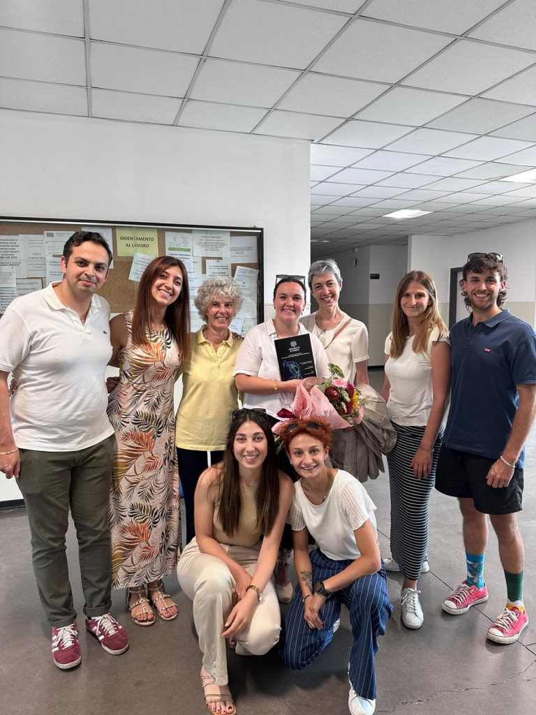
On July 22nd, Ines Parente (ESR13 of the EU H2020 MSCA ITN COLOTAN) performed the defence of her doctoral thesis entitled “Colonoids and cytokines: an in vitro model of drug absorption in the inflamed gut”: she presented her project to prof. Barocelli, prof. Fornai and prof. Colucci, members of the committtee, who had the possibility to appreciate her progresses during the PhD experience, her mastery of the field, and her communicative and self-confident way to share the results of her research. In the end, her final exam was judged as outstanding cum laude. Congratulation, Ines!!!
Abstract
Introduction and Aim
Despite advances in the diagnosis and treatment of inflammatory bowel diseases (IBD), the incidence and prevalence continue to grow. Given the lack of human preclinical studies, reliable models that mimic the inflamed intestinal epithelium are increasingly explored. Patient-derived colonoids are a powerful tool that recapitulate many physiological features of the human colon in vitro. The aim of the present work was to establish an IBD in vitro model based on human colonoid-derived monolayers.
Methods
Colonic crypts, isolated from biopsies of healthy volunteers, were embedded in 70% Matrigel® and grown for 5-7 days. IntestiCult™ OGM was used to generate cystic-like colonoids, further expanded in suspension cultures. After dissociation, cells were seeded at 7.5×10⁵ cells·cm-2 onto 24-well inserts coated with Matrigel® (1:50). The integrity of the epithelial barriers was monitored through TEER measurements. When confluence was reached (TEER ³ 100 W×cm2), IntestiCult™ OGM was switched to ODM. Differentiated colonoid-derived monolayers (CDMs) were exposed for 24 hours to 25 ng·mL-1 TNF-a and 25 ng·mL-1 IFN-g. Changes in cell morphology and viability, IL-8, CCL20, and NF-kB p65 levels, and FITC-D4 apparent permeability (Papp) were evaluated. The Papp values of conventional compounds were determined.
Results
Over 5-15 days, CDMs showed increasing TEER values, confirming the gradual development of epithelial barriers and the strengthening of tight junctions. Immunocytochemical (ICC) staining for villin and MUC2 showed that the cells were polarised and differentiated. Exposure to TNF-a and IFN-g, although heterogeneously, impaired significantly the barrier integrity of CDMs and increased the secretion of IL-8 and CCL20. When the barrier damage was more severe, FITC-D4 Papp and NF-kB p65 levels were also increased, while cell viability was reduced. ICC images showed a large number of apoptotic cells near the basal side, and a decrease in the expression of villin and MUC2, suggesting that cell polarity and differentiation were impaired. While the steady-state flux and the Papp of atenolol increased significantly under inflammatory conditions, the transport of propranolol and budesonide was not affected.
Conclusions
Exposure of CDMs to an inflammatory milieu altered cell morphology, compromised the epithelial barrier integrity, and increased chemotactic cytokine secretion, although to different extents in different patients. Inflammatory conditions significantly increased the paracellular transport of low permeable compounds, highlighting their damaging effect on intercellular proteins.


